Echocardiography
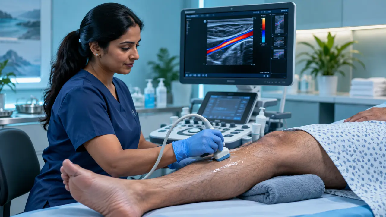
An echocardiogram uses ultrasound to create detailed, real-time images of the heart, allowing Dr. Md. Nagib Mahfuz Khan to assess heart structure, valve function, and pumping efficiency without any radiation or discomfort.
When It Is Recommended
- Breathlessness, chest pain, or unexplained fatigue
- A heart murmur detected on examination
- Follow-up after a heart attack or heart surgery
- Monitoring known valve disease or heart failure
- Routine cardiac risk assessment
What the Test Shows
Echocardiography evaluates the size and thickness of the heart chambers, the pumping strength (ejection fraction), valve function, and blood flow patterns — providing a complete picture of overall heart health.
The Procedure
- Non-invasive and painless, performed with an ultrasound probe on the chest
- Takes around 20-30 minutes to complete
- No special preparation or fasting required
- Results are available for discussion during the same visit
Echocardiography is often the first and most important test used to guide further cardiac management.
Who Should Consult
- New or worsening breathlessness or chest discomfort
- A heart murmur or abnormal heart sound noted by a doctor
- Existing heart disease requiring periodic monitoring
- Pre-operative cardiac assessment before surgery
Frequently Asked Questions
Is echocardiography safe?
Yes, it uses only ultrasound waves with no radiation, making it completely safe and repeatable as often as needed.
Do I need to prepare for the test?
No special preparation is needed — you can eat, drink, and take your regular medication before the test.
How is it different from an ECG?
An ECG records the heart’s electrical activity, while an echocardiogram shows the heart’s actual structure and pumping function through imaging.
Medical content review: Dr. Md. Nagib Mahfuz Khan, BMDC Reg. A-61144. Last updated: August 15, 2026. This information is educational and does not replace an individual medical consultation.